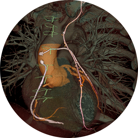
Chest imaging procedures are typically designed to assess cancer, toxin exposure, pulmonary abnormalities, embolism, inflammation, bronchial blockage, pleural effusion, angina, and other disorders.
Some Chest Imaging Procedures Include:
X-ray is the oldest and most economical form of medical imaging. During the procedure, radiation passes through the body onto “film” (now digitized and displayed on a computer screen). In neuroimaging, spinal X-rays are used to assess for the degree of spinal motion with flexion or extension.
Lung cancer CT screening is one of the most accurate diagnostic tools for finding lung cancer at an early stage, when it is most treatable. CT scans of the lung are able to detect small abnormalities in the lungs that could be the beginning stages of lung cancer. These indicators are often not visible on a routine chest X-ray. Since a CT lung screening offers the best opportunity for successful treatment of lung cancer before symptoms are noticed, more physicians are opting for lung cancer screening based on risk factors (like smoking and family history), rather than symptoms.
A ventilation/perfusion lung scan (also known as a V/Q lung scan) assesses the circulation of air (ventilation) and blood (perfusion) in a patient’s lungs. The exam is useful for identifying blood clots or abnormal flow in the lungs or serious lung disorders such as chronic obstructive pulmonary disease (COPD) or a pulmonary embolism. The V/Q scan uses intravenous material (radiotracers) to show the functional health of the organs.


